Ossifying fibroid is a tumor proliferation at the expense of maxillary bones belonging to the complex group of non-odontogenic tumors, having membranous ossification. It accounts for 2.5% of bone tumors and 7% of benign tumors of the head and neck. The objective was to report our diagnostiwc and therapeutic experience through a clinical observation case and literature review. It is a patient of a 7-year-old boy, in the ENT department of the Gabriel Toure University Hospital for painful swelling of the right hemiface associated with a noticeable asymmetry of the face evolving for 04 years. Physical examination revealed a swelling of the right hemiface, extending from the ipsilateral jugal and zygomatic region to the suborbital region, of hard consistency, fixed in relation to the superficial planes and deep, not hot measuring 10 cm over its large diameter, painful with a bulge of the hard ipsilateral palate and a reduction in the lumen of the right nasal cavity. Maxillofacial CT showed a very limited bone density mass that did not take the contrast medium. The histopathological examination after right partial maxillectomy under GA was in favor of FO. Postoperative follow-up was favorable after 04 years of follow-up without recurrence.
This is an Open Access article, distributed under the terms of the Creative Commons Attribution 4.0 International License (http://creativecommons.org/licenses/by/4.0/), which permits unrestricted use, distribution and reproduction in any medium or format, provided the original work is properly cited.
Ossifying fibroid is a tumor proliferation at the expense of maxillary bones belonging to the complex group of non-odontogenic tumors, having membranous ossification
[1]
Julie Guillet, Rémi Curien, François Maschino, Daniel Viennet. Ossifying fibroids of the jaws: diagnostic pitfalls. Med Buccale Chir Buccale, 20 2 (2014) 129-134,
Sheikhi M, Mosavat F, Jalalian F, Rashidipoor R. Central cementifying fibroma of maxilla. Dent Res J (Isfahan) 2013; 10: 122-25.
[1, 2]
. It is a very limited, sometimes even encapsulated, neoformation consisting of fibrous tissue containing variable amounts of calcified material resembling bone and/or cementum
[3]
Beust L, Godey B, Michel M, e Beust L, Godey B, Michel M, et al. Fibrous dysplasias and fibroids ossifying the face, Rev Soc Fr ORL 1997.
[3]
. It accounts for 2.5% of bone tumors and 7% of benign tumors of the head and neck
[4]
Trijolet JP, Permentier J, Sury F, Gogo D, Mejean N, Laure B. Cementoossifying fibroma of the mandible. Eur Ann Otorhinolaryngologol Head Neck Dis 2011; 128: 30-3.
[4]
. It almost exclusively affects the bones of the maxillofacial skeleton
[5]
Eboungabeka Trigo Edith Rose Marcelle, Moussa Mahamane, Bancolé Pognon Sylvie Arlette, Babacar Tamba. The Ossifying Fibroid of the Maxillae: About a Health Sci. Say: Vol 22 (1) January 2021 pp 111-114.
[6]
Prisca A, Hery S, Wilma A, Yudi S. Challenges in managing ossifying fibroma of the maxilla: A case report. Ro Med J. 2024; 71(3).
[5, 6]
. The preferred sites are the mandible followed by the premolar-molar region
[7]
Fujimoto Y, Katoh M, Kawai T, Morita M., Cystic cemento-ossifying fibroma of the petromastoidal region: case report and review of literature. J laryngol otol, 1987; 101: 946-52.
[7]
or the incisal sector for other authors
[8]
Da Silveira, Daniel Trivelato et al. "Ossifying fibroma: report on a clinical case, with the imaging and histopathological diagnosis made and treatment administered." Revista brasileira de ortopedia, vol. 51, 1, 100-4. 21 Dec. 2015,
William H K, Manghan C, Speight M., Juvenile ossifying fibroma. Analysis of eight cases and comparison with other fibro-osseous lesions. J Oral Pathol Med 2000; 29: 13-8.
[8, 9]
. It usually occurs between the second and fourth decade, with a male/female ratio of 1:5. Slow-growing, normally well-defined and asymptomatic, but over time the lesion can become quite large and cause facial deformity
[10]
Javier Silvestre-Rangil, Francisco Javier Silvestre., Cemento-ossifying fibroma of the mandible. J Clin Exp Dent. 2011; 3(1): 66-9.
[10]
.
Because of the aggressive characteristics and high potential for recurrence of FOJ, the standard treatment is complete excision with a margin of safety. Therefore, a complete resection could prove difficult in terms of reconstructing the continuity defect, both aesthetically and functionally
[11]
Sanchis JM, Peñarrocha M, Balaguer JM, Camacho F., Cemento-ossifying mandibular a presentation of two cases and review of the literature. Med Oral 2004; 9: 69-73.
[11]
.
The delay in consultation and the attribution of these tumors to mystical factors increase their severity, thus making their management difficult and clouding the prognosis. This justifies our clinical presentation.
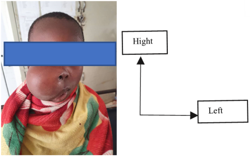
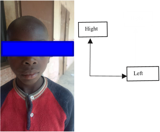
Figure 1. Anterior view of the face preoperatively showing a right jugal swelling.
This is a 7-year-old male patient residing in Bougouni, who was referred to the CSREF of Bougouni for painful swelling of the right hemiface associated with a significant asymmetry of the face that has been evolving for 4 years. The patient is in good general health and no medical history is reported. Examination of the cervicofacial skin reveals: a swelling of the right hemiface, extending from the ipsilateral jugal and zygomatic region to the suborbital region with notable asymmetry of the face, of hard consistency, fixed in relation to the superficial planes and deep, not hot measuring 10 cm on its large diameter, not painful on palpation. The skin in look healthy. On examination of the oral cavity and the oropharynx, a bulge of the hard palate was noted ipsilateral. On rhinoscopy, there was a reduction in the lumen of the right nasal cavity. The rest of the physical examination was normal.
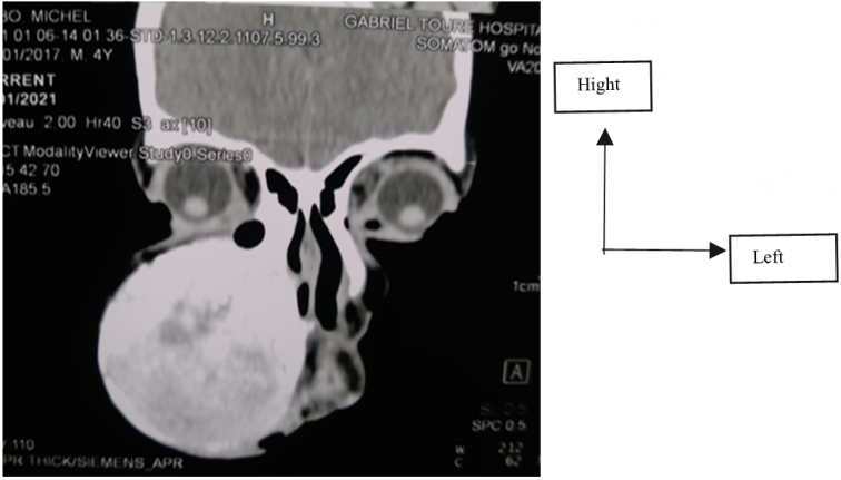
Figure 3. Coronal section CT image showing the hyperdense mass in the maxillary sinus with mass effect on the ipsilateral nasal cavity.
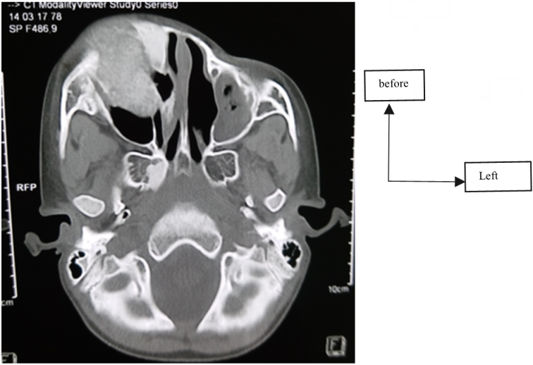
On maxillofacial computed tomography, we have demonstrated a mass of well-limited bone density of the anterolateral bone wall of the right maxillary sinus, with regular contours, extending to the ipsilateral orbital floor and not taking the contrast medium. The patient's blood operability and chest X-ray tests returned normal.
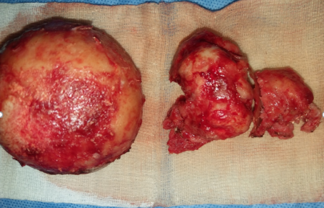
A right partial maxillectomy was performed under general anesthesia after a gingivo-vestibular incision extended to the right corner of the lip for about 5 cm. There was no reconstruction of the right jaw by autologous grafts, nor extraction of the dental alveoli.
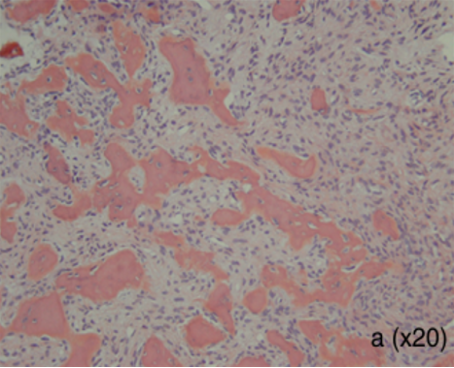
The histopathological examination of the operative specimen revealed a benign tumor lesion of a fibroosseous nature; bone structures of variable size observed within a fairly high cellularity contingent, spindle-shaped, and devoid of atypicality, in favor of the ossifying fibroid.
Figure 5. Histological section showing numerous spindle cells, globular fibrillar bone structures, without a clear osteoblastic border.
The postoperative follow-up of the patient under antibiotic prophylaxis, corticosteroid therapy, analgesics and nasal lavage (after diseming), was favorable after 04 years of follow-up without recurrence.
Ossifying fibroid is a benign fibroosseous lesion of the jawbones containing varying amounts of calcified deposits of bone, cementum or a mixture of both. Taking into account the amount of hard tissue formed in the lesion, fibroid ossifying has been subdivided histologically into fibroid ossifying and fibroid cementifying. Included in odontogenic tumors for cementifying fibroid and non-odontogenic bone tumors for ossifying fibroid, respectively.
It usually occurs between the second and fourth decade, with a male/female ratio of 1:5. This female predominance is commonly accepted, even if no study on a large number of cases has been conducted so far
[12]
Kadiri F, Laraqui NZ, Touhani M, Benghale M A, Mokrim B, Chekkoury-Idrisi A, Bencha Kroun Y., Ossifying fibroids of the maxilla., Rev Laryngol 1993; 114: 349-53.
[12]
. Despite these data from the literature, our observation reports an unusual case in a 7-year-old male child.
The circumstances in which ossifying fibroids were discovered may be fortuitous because its evolution is slow and can remain asymptomatic for a long time. In an advanced stage, it causes functional and aesthetic discomfort, facial asymmetry, gingival swelling, mobility or tooth displacement
[10]
Javier Silvestre-Rangil, Francisco Javier Silvestre., Cemento-ossifying fibroma of the mandible. J Clin Exp Dent. 2011; 3(1): 66-9.
[10]
. The maxillofacial localization of the F.O. is preferentially mandibular in 75% of cases, involving the premolar-molar region
[10]
Javier Silvestre-Rangil, Francisco Javier Silvestre., Cemento-ossifying fibroma of the mandible. J Clin Exp Dent. 2011; 3(1): 66-9.
[10]
. The maxilla can be affected particularly in the anterior sector
[13]
Barnes L, Eveson J W, Reichart P, D. Sidransky. Ranking the World Tumor Health Organization, The Editors of Head and Neck Tumors. Lyon: IARC press; 2005. Pathology and genetics.
[14]
Burns J, Le Zzoni J, Reibel J. Pathologic quiz box 2. Arch Otolaryngol Head Neck Surg 1996; 122: 681-33.
[13, 14]
, as in our observation.
The evolution of this tumor is constantly benign, however if left untreated, the tumor grows slowly and can reach a considerable volume with rupture of the bony cortex and diffusion into the soft tissues.
From a radiological point of view, the ossifying fibroid is expressed on CT by a well-defined osteolytic image of unilocular or multilocular, associated or not with root resorptions
[15]
Diniz JA, Siqueira ADS, Araujo GM, Faro TF, Torres LHS, Oliveira E Silva ED, et al. Intraoral approach for surgical treatment of psammomatoid juvenile ossifying fibroma. J Cranio-fac Surg 2020; 31: 306-9.
Gantala R, Vemula AY, Kubbi JR, Sekhar MM, Jhawar D. Psammomatoid juvenile ossifying fibroma involving upper jaw: a rare case report. J Clin Diagn Res 2015; 9: ZD17-9.
[15, 16]
, consistent with our case.
Clinical and radiological examinations alone do not provide sufficient diagnostic arguments, only comparison with histopathological examination makes it possible to specify the exact nature of the tumor lesions.
The standard treatment is complete excision with a margin of safety due to the aggressive characteristics and potential for recurrence of some clinical forms of fibroid ossifican. In the case of large tumors, large incisions are necessary to expose the tumor
[17]
Wong WW, Martin MC. Reconstruction of extended orbitomaxillectomy and hemimandibulectomy defects with fibula flaps and patient-specific implants. J Craniofac Surg 2016; 27: 380-4.
[17]
as was the case for our observation. Therefore, a complete resection could prove difficult in terms of reconstructing the continuity defect, both aesthetically and functionally. For small tumors, minimally invasive surgery may be possible via smaller incisions using an intraoral approach or endonasal endoscopy
[15]
Diniz JA, Siqueira ADS, Araujo GM, Faro TF, Torres LHS, Oliveira E Silva ED, et al. Intraoral approach for surgical treatment of psammomatoid juvenile ossifying fibroma. J Cranio-fac Surg 2020; 31: 306-9.
Gantala R, Vemula AY, Kubbi JR, Sekhar MM, Jhawar D. Psammomatoid juvenile ossifying fibroma involving upper jaw: a rare case report. J Clin Diagn Res 2015; 9: ZD17-9.
[18]
Rizk Saad H, M. Kamal N and W. Amer H. Case Report: rare hybrid lesion of a central giant cell granuloma within a juvenile ossifying fibroma [version 1; peer review: 1 approved, 2 approved with reservations] F1000 Research 2019, 8: 1218
Although there is no consensus on the optimal method or timing of reconstruction of postoperative defects, autologous bone grafts are considered the standard gold, with or without the use of a microsurgical technique
[17]
Wong WW, Martin MC. Reconstruction of extended orbitomaxillectomy and hemimandibulectomy defects with fibula flaps and patient-specific implants. J Craniofac Surg 2016; 27: 380-4.
[17]
.
Recurrences of F.O are reported at varying rates: 10 to 28% after enucleation and less than (5%) after excision, which justifies clinical and radiological monitoring over several years
[14]
Burns J, Le Zzoni J, Reibel J. Pathologic quiz box 2. Arch Otolaryngol Head Neck Surg 1996; 122: 681-33.
[19]
Damjanon I, Linder J. Anderson's pathology (pp 1606-1607). Mosby, St Louis, 1996. P. 1606-7.
[14, 19]
. Malignant transformations of the fibroid ossifying are rare
[18]
Rizk Saad H, M. Kamal N and W. Amer H. Case Report: rare hybrid lesion of a central giant cell granuloma within a juvenile ossifying fibroma [version 1; peer review: 1 approved, 2 approved with reservations] F1000 Research 2019, 8: 1218
Fibroosseous lesions of the facial mass remain rare entities, the diagnosis of which remains difficult both histologically and radiologically. The totality (comparison) of the clinical, histological and radiological data is often necessary to make the diagnosis. The treatment is based on surgery, the indications of which are dictated by the age of the patient, the volume and location of the tumor, and the possible functional or aesthetic impact.
Abbreviations
FO
Fibroid Ossifying
CSREF
Reference Health Centre
ENT-CCF
Otorhinolaryngology and Head and Neck Surgery
CT scan
Computed Tomography
FOJ
Juvenile Fibroid Ossificans
Funding
The authors state that they have not received any specific funding for this work.
Conflicts of Interest
The authors declare no conflicts of interest.
References
[1]
Julie Guillet, Rémi Curien, François Maschino, Daniel Viennet. Ossifying fibroids of the jaws: diagnostic pitfalls. Med Buccale Chir Buccale, 20 2 (2014) 129-134,
Sheikhi M, Mosavat F, Jalalian F, Rashidipoor R. Central cementifying fibroma of maxilla. Dent Res J (Isfahan) 2013; 10: 122-25.
[3]
Beust L, Godey B, Michel M, e Beust L, Godey B, Michel M, et al. Fibrous dysplasias and fibroids ossifying the face, Rev Soc Fr ORL 1997.
[4]
Trijolet JP, Permentier J, Sury F, Gogo D, Mejean N, Laure B. Cementoossifying fibroma of the mandible. Eur Ann Otorhinolaryngologol Head Neck Dis 2011; 128: 30-3.
[5]
Eboungabeka Trigo Edith Rose Marcelle, Moussa Mahamane, Bancolé Pognon Sylvie Arlette, Babacar Tamba. The Ossifying Fibroid of the Maxillae: About a Health Sci. Say: Vol 22 (1) January 2021 pp 111-114.
[6]
Prisca A, Hery S, Wilma A, Yudi S. Challenges in managing ossifying fibroma of the maxilla: A case report. Ro Med J. 2024; 71(3).
[7]
Fujimoto Y, Katoh M, Kawai T, Morita M., Cystic cemento-ossifying fibroma of the petromastoidal region: case report and review of literature. J laryngol otol, 1987; 101: 946-52.
[8]
Da Silveira, Daniel Trivelato et al. "Ossifying fibroma: report on a clinical case, with the imaging and histopathological diagnosis made and treatment administered." Revista brasileira de ortopedia, vol. 51, 1, 100-4. 21 Dec. 2015,
William H K, Manghan C, Speight M., Juvenile ossifying fibroma. Analysis of eight cases and comparison with other fibro-osseous lesions. J Oral Pathol Med 2000; 29: 13-8.
[10]
Javier Silvestre-Rangil, Francisco Javier Silvestre., Cemento-ossifying fibroma of the mandible. J Clin Exp Dent. 2011; 3(1): 66-9.
[11]
Sanchis JM, Peñarrocha M, Balaguer JM, Camacho F., Cemento-ossifying mandibular a presentation of two cases and review of the literature. Med Oral 2004; 9: 69-73.
[12]
Kadiri F, Laraqui NZ, Touhani M, Benghale M A, Mokrim B, Chekkoury-Idrisi A, Bencha Kroun Y., Ossifying fibroids of the maxilla., Rev Laryngol 1993; 114: 349-53.
[13]
Barnes L, Eveson J W, Reichart P, D. Sidransky. Ranking the World Tumor Health Organization, The Editors of Head and Neck Tumors. Lyon: IARC press; 2005. Pathology and genetics.
[14]
Burns J, Le Zzoni J, Reibel J. Pathologic quiz box 2. Arch Otolaryngol Head Neck Surg 1996; 122: 681-33.
[15]
Diniz JA, Siqueira ADS, Araujo GM, Faro TF, Torres LHS, Oliveira E Silva ED, et al. Intraoral approach for surgical treatment of psammomatoid juvenile ossifying fibroma. J Cranio-fac Surg 2020; 31: 306-9.
Gantala R, Vemula AY, Kubbi JR, Sekhar MM, Jhawar D. Psammomatoid juvenile ossifying fibroma involving upper jaw: a rare case report. J Clin Diagn Res 2015; 9: ZD17-9.
[17]
Wong WW, Martin MC. Reconstruction of extended orbitomaxillectomy and hemimandibulectomy defects with fibula flaps and patient-specific implants. J Craniofac Surg 2016; 27: 380-4.
[18]
Rizk Saad H, M. Kamal N and W. Amer H. Case Report: rare hybrid lesion of a central giant cell granuloma within a juvenile ossifying fibroma [version 1; peer review: 1 approved, 2 approved with reservations] F1000 Research 2019, 8: 1218
Moussa, K., Ibrahim, D., Tata, T., Oumar, K., Aissata, O., et al. (2025). An Unusual Case of Fibroid Ossifying of the Maxilla in a Male Child. Clinical Medicine Research, 14(5), 194-197. https://doi.org/10.11648/j.cmr.20251405.15
Moussa, K.; Ibrahim, D.; Tata, T.; Oumar, K.; Aissata, O., et al. An Unusual Case of Fibroid Ossifying of the Maxilla in a Male Child. Clin. Med. Res.2025, 14(5), 194-197. doi: 10.11648/j.cmr.20251405.15
Moussa K, Ibrahim D, Tata T, Oumar K, Aissata O, et al. An Unusual Case of Fibroid Ossifying of the Maxilla in a Male Child. Clin Med Res. 2025;14(5):194-197. doi: 10.11648/j.cmr.20251405.15
@article{10.11648/j.cmr.20251405.15,
  author = {Konate Moussa and Dicko Ibrahim and Toure Tata and Konate Oumar and Ouane Aissata and Coulibaly Assitan Kole and Tangara Mariam and Traore Nouhoum and Simpara Gaoussou and Keita Moussa Bourama and Berthe Ismael and Cisse Naouma and Konate N'faly and Diarra Kassim and Kone Fatogoma Issa and Guindo Boubacary and Soumaoro Siaka and Singare Kadidiatou and Keita Mohamed Amadou},
  title = {An Unusual Case of Fibroid Ossifying of the Maxilla in a Male Child},
  journal = {Clinical Medicine Research},
  volume = {14},
  number = {5},
  pages = {194-197},
  doi = {10.11648/j.cmr.20251405.15},
  url = {https://doi.org/10.11648/j.cmr.20251405.15},
  eprint = {https://article.sciencepublishinggroup.com/pdf/10.11648.j.cmr.20251405.15},
  abstract = {Ossifying fibroid is a tumor proliferation at the expense of maxillary bones belonging to the complex group of non-odontogenic tumors, having membranous ossification. It accounts for 2.5% of bone tumors and 7% of benign tumors of the head and neck. The objective was to report our diagnostiwc and therapeutic experience through a clinical observation case and literature review. It is a patient of a 7-year-old boy, in the ENT department of the Gabriel Toure University Hospital for painful swelling of the right hemiface associated with a noticeable asymmetry of the face evolving for 04 years. Physical examination revealed a swelling of the right hemiface, extending from the ipsilateral jugal and zygomatic region to the suborbital region, of hard consistency, fixed in relation to the superficial planes and deep, not hot measuring 10 cm over its large diameter, painful with a bulge of the hard ipsilateral palate and a reduction in the lumen of the right nasal cavity. Maxillofacial CT showed a very limited bone density mass that did not take the contrast medium. The histopathological examination after right partial maxillectomy under GA was in favor of FO. Postoperative follow-up was favorable after 04 years of follow-up without recurrence.
},
 year = {2025}
}
TY  - JOUR
T1  - An Unusual Case of Fibroid Ossifying of the Maxilla in a Male Child
AU  - Konate Moussa
AU  - Dicko Ibrahim
AU  - Toure Tata
AU  - Konate Oumar
AU  - Ouane Aissata
AU  - Coulibaly Assitan Kole
AU  - Tangara Mariam
AU  - Traore Nouhoum
AU  - Simpara Gaoussou
AU  - Keita Moussa Bourama
AU  - Berthe Ismael
AU  - Cisse Naouma
AU  - Konate N'faly
AU  - Diarra Kassim
AU  - Kone Fatogoma Issa
AU  - Guindo Boubacary
AU  - Soumaoro Siaka
AU  - Singare Kadidiatou
AU  - Keita Mohamed Amadou
Y1  - 2025/10/10
PY  - 2025
N1  - https://doi.org/10.11648/j.cmr.20251405.15
DO  - 10.11648/j.cmr.20251405.15
T2  - Clinical Medicine Research
JF  - Clinical Medicine Research
JO  - Clinical Medicine Research
SP  - 194
EP  - 197
PB  - Science Publishing Group
SN  - 2326-9057
UR  - https://doi.org/10.11648/j.cmr.20251405.15
AB  - Ossifying fibroid is a tumor proliferation at the expense of maxillary bones belonging to the complex group of non-odontogenic tumors, having membranous ossification. It accounts for 2.5% of bone tumors and 7% of benign tumors of the head and neck. The objective was to report our diagnostiwc and therapeutic experience through a clinical observation case and literature review. It is a patient of a 7-year-old boy, in the ENT department of the Gabriel Toure University Hospital for painful swelling of the right hemiface associated with a noticeable asymmetry of the face evolving for 04 years. Physical examination revealed a swelling of the right hemiface, extending from the ipsilateral jugal and zygomatic region to the suborbital region, of hard consistency, fixed in relation to the superficial planes and deep, not hot measuring 10 cm over its large diameter, painful with a bulge of the hard ipsilateral palate and a reduction in the lumen of the right nasal cavity. Maxillofacial CT showed a very limited bone density mass that did not take the contrast medium. The histopathological examination after right partial maxillectomy under GA was in favor of FO. Postoperative follow-up was favorable after 04 years of follow-up without recurrence.

VL  - 14
IS  - 5
ER  -
Department of ENT (Otorhinolaryngology) and Head and Neck Surgery, Gabriel Toure University Hospital, Bamako, Mali
Coulibaly Assitan Kole
Department of ENT (Otorhinolaryngology) and Head and Neck Surgery, Gabriel Toure University Hospital, Bamako, Mali
Tangara Mariam
Department of ENT (Otorhinolaryngology) and Head and Neck Surgery, Gabriel Toure University Hospital, Bamako, Mali
Traore Nouhoum
Department of ENT (Otorhinolaryngology) and Head and Neck Surgery, Gabriel Toure University Hospital, Bamako, Mali
Simpara Gaoussou
Department of ENT (Otorhinolaryngology) and Head and Neck Surgery, Gabriel Toure University Hospital, Bamako, Mali
Keita Moussa Bourama
Department of ENT (Otorhinolaryngology) and Head and Neck Surgery, Gabriel Toure University Hospital, Bamako, Mali
Berthe Ismael
Department of ENT (Otorhinolaryngology) and Head and Neck Surgery, Gabriel Toure University Hospital, Bamako, Mali
Cisse Naouma
Department of ENT (Otorhinolaryngology) and Head and Neck Surgery, Gabriel Toure University Hospital, Bamako, Mali
Konate N'faly
Department of ENT (Otorhinolaryngology) and Head and Neck Surgery, Gabriel Toure University Hospital, Bamako, Mali
Diarra Kassim
Department of ENT (Otorhinolaryngology) and Head and Neck Surgery, Gabriel Toure University Hospital, Bamako, Mali
Kone Fatogoma Issa
Department of ENT (Otorhinolaryngology) and Head and Neck Surgery, Gabriel Toure University Hospital, Bamako, Mali; Faculty of Medicine and Odontostomatology, USTTB (Université des Sciences, des Techniques et des Technologies de Bamako), Bamako, Mali
Department of ENT (Otorhinolaryngology) and Head and Neck Surgery, Gabriel Toure University Hospital, Bamako, Mali; Faculty of Medicine and Odontostomatology, USTTB (Université des Sciences, des Techniques et des Technologies de Bamako), Bamako, Mali
Soumaoro Siaka
Department of ENT (Otorhinolaryngology) and Head and Neck Surgery, Gabriel Toure University Hospital, Bamako, Mali; Faculty of Medicine and Odontostomatology, USTTB (Université des Sciences, des Techniques et des Technologies de Bamako), Bamako, Mali
Singare Kadidiatou
Department of ENT (Otorhinolaryngology) and Head and Neck Surgery, Gabriel Toure University Hospital, Bamako, Mali; Faculty of Medicine and Odontostomatology, USTTB (Université des Sciences, des Techniques et des Technologies de Bamako), Bamako, Mali
Keita Mohamed Amadou
Department of ENT (Otorhinolaryngology) and Head and Neck Surgery, Gabriel Toure University Hospital, Bamako, Mali; Faculty of Medicine and Odontostomatology, USTTB (Université des Sciences, des Techniques et des Technologies de Bamako), Bamako, Mali
Moussa, K., Ibrahim, D., Tata, T., Oumar, K., Aissata, O., et al. (2025). An Unusual Case of Fibroid Ossifying of the Maxilla in a Male Child. Clinical Medicine Research, 14(5), 194-197. https://doi.org/10.11648/j.cmr.20251405.15
Moussa, K.; Ibrahim, D.; Tata, T.; Oumar, K.; Aissata, O., et al. An Unusual Case of Fibroid Ossifying of the Maxilla in a Male Child. Clin. Med. Res.2025, 14(5), 194-197. doi: 10.11648/j.cmr.20251405.15
Moussa K, Ibrahim D, Tata T, Oumar K, Aissata O, et al. An Unusual Case of Fibroid Ossifying of the Maxilla in a Male Child. Clin Med Res. 2025;14(5):194-197. doi: 10.11648/j.cmr.20251405.15
@article{10.11648/j.cmr.20251405.15,
  author = {Konate Moussa and Dicko Ibrahim and Toure Tata and Konate Oumar and Ouane Aissata and Coulibaly Assitan Kole and Tangara Mariam and Traore Nouhoum and Simpara Gaoussou and Keita Moussa Bourama and Berthe Ismael and Cisse Naouma and Konate N'faly and Diarra Kassim and Kone Fatogoma Issa and Guindo Boubacary and Soumaoro Siaka and Singare Kadidiatou and Keita Mohamed Amadou},
  title = {An Unusual Case of Fibroid Ossifying of the Maxilla in a Male Child},
  journal = {Clinical Medicine Research},
  volume = {14},
  number = {5},
  pages = {194-197},
  doi = {10.11648/j.cmr.20251405.15},
  url = {https://doi.org/10.11648/j.cmr.20251405.15},
  eprint = {https://article.sciencepublishinggroup.com/pdf/10.11648.j.cmr.20251405.15},
  abstract = {Ossifying fibroid is a tumor proliferation at the expense of maxillary bones belonging to the complex group of non-odontogenic tumors, having membranous ossification. It accounts for 2.5% of bone tumors and 7% of benign tumors of the head and neck. The objective was to report our diagnostiwc and therapeutic experience through a clinical observation case and literature review. It is a patient of a 7-year-old boy, in the ENT department of the Gabriel Toure University Hospital for painful swelling of the right hemiface associated with a noticeable asymmetry of the face evolving for 04 years. Physical examination revealed a swelling of the right hemiface, extending from the ipsilateral jugal and zygomatic region to the suborbital region, of hard consistency, fixed in relation to the superficial planes and deep, not hot measuring 10 cm over its large diameter, painful with a bulge of the hard ipsilateral palate and a reduction in the lumen of the right nasal cavity. Maxillofacial CT showed a very limited bone density mass that did not take the contrast medium. The histopathological examination after right partial maxillectomy under GA was in favor of FO. Postoperative follow-up was favorable after 04 years of follow-up without recurrence.
},
 year = {2025}
}
TY  - JOUR
T1  - An Unusual Case of Fibroid Ossifying of the Maxilla in a Male Child
AU  - Konate Moussa
AU  - Dicko Ibrahim
AU  - Toure Tata
AU  - Konate Oumar
AU  - Ouane Aissata
AU  - Coulibaly Assitan Kole
AU  - Tangara Mariam
AU  - Traore Nouhoum
AU  - Simpara Gaoussou
AU  - Keita Moussa Bourama
AU  - Berthe Ismael
AU  - Cisse Naouma
AU  - Konate N'faly
AU  - Diarra Kassim
AU  - Kone Fatogoma Issa
AU  - Guindo Boubacary
AU  - Soumaoro Siaka
AU  - Singare Kadidiatou
AU  - Keita Mohamed Amadou
Y1  - 2025/10/10
PY  - 2025
N1  - https://doi.org/10.11648/j.cmr.20251405.15
DO  - 10.11648/j.cmr.20251405.15
T2  - Clinical Medicine Research
JF  - Clinical Medicine Research
JO  - Clinical Medicine Research
SP  - 194
EP  - 197
PB  - Science Publishing Group
SN  - 2326-9057
UR  - https://doi.org/10.11648/j.cmr.20251405.15
AB  - Ossifying fibroid is a tumor proliferation at the expense of maxillary bones belonging to the complex group of non-odontogenic tumors, having membranous ossification. It accounts for 2.5% of bone tumors and 7% of benign tumors of the head and neck. The objective was to report our diagnostiwc and therapeutic experience through a clinical observation case and literature review. It is a patient of a 7-year-old boy, in the ENT department of the Gabriel Toure University Hospital for painful swelling of the right hemiface associated with a noticeable asymmetry of the face evolving for 04 years. Physical examination revealed a swelling of the right hemiface, extending from the ipsilateral jugal and zygomatic region to the suborbital region, of hard consistency, fixed in relation to the superficial planes and deep, not hot measuring 10 cm over its large diameter, painful with a bulge of the hard ipsilateral palate and a reduction in the lumen of the right nasal cavity. Maxillofacial CT showed a very limited bone density mass that did not take the contrast medium. The histopathological examination after right partial maxillectomy under GA was in favor of FO. Postoperative follow-up was favorable after 04 years of follow-up without recurrence.

VL  - 14
IS  - 5
ER  -